
Introduction to Tuberculosis
Tuberculosis, often called TB, is a serious infection caused by bacteria. It mainly affects the lungs but can also spread to other parts of the body. According to the World Health Organization (WHO), millions of people worldwide are affected by TB each year. In many places, including cities with high population density, TB remains a common health concern. Early detection is important for treatment and prevention.
Importance of Chest X-rays in TB Diagnosis
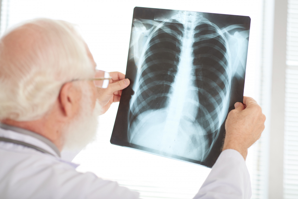
Doctors often use chest X-rays to help diagnose tuberculosis. While a chest X-ray cannot confirm TB alone, it gives important clues. For example, it can show changes in the lungs that suggest infection. Because TB can look like other lung problems, doctors use X-rays along with other tests. In many countries, chest X-rays are a first step when TB is suspected.
Common Chest X-ray Findings in Tuberculosis
Chest X-ray findings in tuberculosis can vary. However, some signs are more common in people with TB. Knowing these signs helps doctors decide on further tests. Here are some typical findings:
Although these findings are common, not everyone with TB will have all of them. Sometimes, the X-ray may look normal even if TB is present.
How Radiologists Interpret TB on X-rays
Radiologists are doctors who read and interpret X-rays. They look for patterns that match signs of tuberculosis on chest X-ray. For example, they check the location and shape of any shadows or cavities. Additionally, they compare the X-ray to previous images if available. This helps them see if the changes are new or old. Because other diseases can look similar, radiologists work closely with your doctor to make the best diagnosis.
What to Expect During a Chest X-ray
Getting a chest X-ray is quick and painless. First, you will stand or sit in front of the X-ray machine. Next, a technician will ask you to take a deep breath and hold it for a few seconds. This helps get a clear picture. The process usually takes less than 10 minutes. You can return to normal activities right after the test. There is only a small amount of radiation used, so the risk is very low.
Limitations of Chest X-rays in TB Detection
While chest X-rays are helpful, they have some limits. For instance, not all TB cases show clear signs on X-ray. Early TB or TB in children may be hard to spot. Also, other lung diseases can look like TB on X-ray. Because of this, doctors often use other tests, such as sputum tests or blood tests, to confirm TB. Therefore, a normal X-ray does not always mean you are free from TB.
Prevention and Next Steps if TB is Suspected
If you have symptoms like a cough lasting more than two weeks, fever, night sweats, or weight loss, you should see a doctor. Early testing is important to prevent the spread of TB. If your doctor suspects TB, they may order a chest X-ray and other tests. In addition, you should:
Prevention also includes good ventilation at home and regular health check-ups, especially in areas where TB is common.
Conclusion
In summary, chest X-ray findings in tuberculosis help doctors spot signs of TB in the lungs. However, X-rays are only one part of the diagnosis. If you or someone you know is experiencing symptoms of tuberculosis, consult a healthcare specialist for personalized advice.